The Evolution of Diagnostic Accuracy in Hand Surgery
Modern orthopedic care relies on more than routine physical assessments to address complex hand and wrist conditions. While initial examinations provide necessary context, they often lack the precision required to visualize subtle nerve lesions, tendon functional integrity, or deep-tissue injuries per PMC10953511. Integrating advanced imaging like magnetic resonance arthrography or dynamic ultrasonography allows specialists to move beyond guesswork and tailor precise intervention strategies.
Achieving high-level diagnostic accuracy requires close collaboration between the surgeon and radiologists. Relying on imaging without clinical context can lead to misinterpretation, as seen in cases where asymptomatic incidental findings appear on high-resolution scans per TandF. As a specialist in Berkeley, Rebecca S. Yu, MD emphasizes that surgeons must clearly define the clinical question to ensure radiologic reports directly inform surgical planning, unlike general practices that may order reflexive, unnecessary testing. This partnership ensures that every diagnostic tool, from advanced CT reconstruction to Point of Care Ultrasound, serves the patient's specific recovery goals.
Foundational Imaging Modalities for Hand and Wrist Care
Diagnostic imaging is fundamental to orthopedic care, providing the detailed visual insights necessary for accurately diagnosing and treating musculoskeletal conditions. By utilizing various modalities, such as X-rays for bone structure, MRI for soft tissue assessment, and CT scans for complex bone visualization, specialists can pinpoint the exact cause of pain. These tools differentiate between overlapping symptoms, allowing for precise identification of issues like fractures, ligament tears, or degenerative changes.
- Conventional radiographs remain the gold standard for initial assessments because they effectively confirm osseous alignment and detect fractures at a relatively low cost.
- Magnetic resonance imaging is preferred for evaluating soft tissue injuries, including cartilage or ligament damage, though it should be used selectively to avoid acting on incidental findings that lack clinical significance.
- Computed tomography provides superior definition for complex, intra-articular fractures. When standard scans are insufficient, MR arthrography serves as a specialized tool to capture higher sensitivity for ligamentous tears like those involving the triangular fibrocartilage complex.
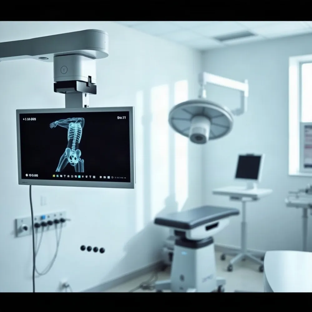
Dr. Rebecca S. Yu integrates these modalities to guide personalized treatment plans. While some clinics rely solely on routine diagnostics, rebeccasyumd.com emphasizes using high-resolution ultrasound and CT with 3D reconstruction to map injuries before entering the OR. This approach allows for a clearer understanding of nerve, tendon, and ligament integrity beyond what static images reveal. By matching the correct modality to the specific clinical question, practitioners avoid unnecessary radiation and costs while optimizing surgical decision-making for every patient.
Selecting the Right Imaging Study for Your Injury
How do I know whether an X-ray, CT scan, or MRI is best for my wrist injury?
Determining the right imaging study depends on the nature of your symptoms and what your doctor suspects may be injured. X-rays are almost always the first step, as they are quick and effective for identifying obvious fractures, dislocations, or major alignment issues in the bones. If your initial X-ray is inconclusive but there is still a high clinical suspicion of an injury, a CT scan or MRI may be necessary to gain a clearer picture. A CT scan is generally preferred if we need highly detailed, 360-degree views of complex bone fractures, whereas an MRI is the gold standard for evaluating soft tissues, such as ligaments, tendons, cartilage, and bone marrow edema. Ultimately, the choice of imaging is a clinical decision based on your specific presentation, which we will evaluate during your examination.
At rebeccasyumd.com, we tailor diagnostic strategies to your unique clinical needs. While standard radiographs are the primary tool for osseous alignment, computed tomography is superior for assessing occult or complex intra-articular fractures where radiographs may remain equivocal. For suspected scaphoid fractures that fail to appear on plain film, advanced imaging is essential to identify osteonecrosis or vascularity issues that dictate your treatment path.
We approach magnetic resonance imaging with precision to avoid the trap of clinical over-interpretation. Because incidental findings such as small ganglions or minor tendon irregularities often correlate poorly with actual pain, we prioritize findings that align strictly with your physical symptoms. By integrating imaging into a focused clinical rehabilitation plan, we ensure that diagnostic data serves your recovery rather than simply adding complexity to your care.
Dynamic Assessment and Ultrasound Potential
Musculoskeletal ultrasound has transformed the diagnostic approach to hand and wrist conditions by offering real-time, dynamic visualization of soft tissues. Unlike static imaging, musculoskeletal ultrasound allows surgeons like Rebecca S. Yu, MD to observe pathology during active movement. This capability is vital for conditions such as trigger finger or tendon subluxation, where the underlying mechanical defect only appears when the hand is in motion.
A primary benefit of this modality is the ability to monitor fine structures like digital nerves and tendon gliding. Ultrahigh frequency ultrasound delivers resolution as fine as 30 micrometers, permitting clinicians to inspect individual nerve fascicles and evaluate pulley integrity. While standard MRIs are useful for static anatomy, they can produce incidental findings that lack clinical relevance, as noted in studies analyzing hundreds of hand and wrist images. In contrast, ultrasound provides direct functional feedback, helping determine if a patient needs tenolysis or nerve grafting.
For patients visiting our clinic in Berkeley, Point of Care Ultrasound (POCUS) serves as a natural extension of the physical examination. This portable technology allows for immediate assessment of soft tissue infections, masses, or foreign bodies, often providing diagnostic answers during the initial consultation. The precision afforded by POCUS extends to procedural guidance as well, where visual confirmation improves the accuracy of joint injections for conditions like carpal tunnel syndrome compared to traditional blind techniques.
| Feature | Clinical Benefit | Outcome |
|---|---|---|
| Dynamic imaging | Real-time movement tracking | Identifies snapping and catching |
| High resolution | Visualization of fascicles | Precise nerve compression mapping |
| POCUS integration | Bedside diagnostic utility | Reduces need for follow-up |
Ultrahigh Frequency and Advanced Specialized Imaging
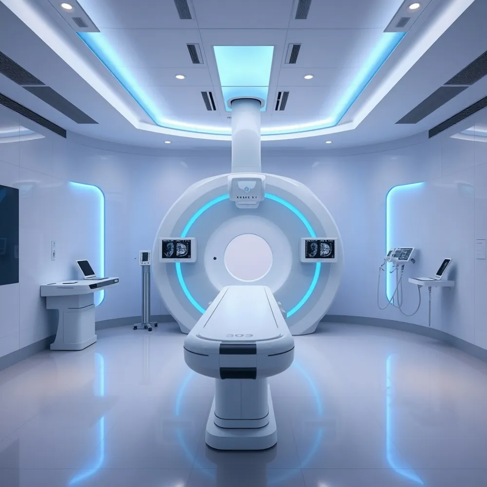 For surgeons at rebeccasyumd.com, achieving diagnostic precision in the hand and wrist often requires moving beyond standard imaging. Ultrahigh frequency ultrasound (UHFUS) has emerged as a high-resolution tool, utilizing frequencies up to 70 MHz to capture images with 30-micrometer resolution. This level of detail allows specialists to visualize individual nerve fascicles and fine superficial structures that conventional ultrasound systems, typically operating between 5 and 12 MHz, often miss.
When assessing complex bone injuries, specialized CT protocols offer distinct advantages over traditional multi-slice tomography. Cone-beam computed tomography (CBCT) provides high spatial resolution while significantly reducing radiation exposure. Unlike conventional CT scanners, CBCT is particularly effective at minimizing metal artifacts, which is critical when evaluating bone healing around previous surgical hardware.
The integration of tomosynthesis into the surgical workflow further enhances precision by providing three-dimensional, scrollable images that outperform standard fluoroscopy. This modality allows for clearer assessment of intra-articular fractures and confirmation of hardware placement without the higher costs associated with full-scale CT scans. At rebeccasyumd.com, these advanced techniques are utilized to refine treatment pathways, ensuring that every diagnostic step informs a highly specific and targeted operative plan.
| Modality | Key Advantage | Clinical Utility |
|---|---|---|
| UHFUS | High resolution | Nerve and tendon imaging |
| CBCT | Low metal artifact | Complex fracture assessment |
| Tomosynthesis | 3D visualization | Hardware verification |
Harnessing Artificial Intelligence for Surgical Precision
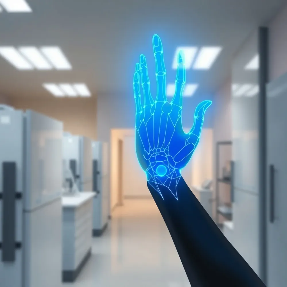 Artificial intelligence is rapidly transforming orthopedic surgery by serving as a powerful tool for diagnostic assistance, surgical planning, and predictive modeling. Recent advancements in [deep learning and convolutional neural networks allow for automated medical image segmentation and 3D modeling, which enables surgeons to move beyond traditional visual limitations.
How AI assists in detecting subtle, occult fractures
Machine learning algorithms applied to X-rays and CT scans can increase the diagnostic accuracy of occult foot and ankle fractures to as high as 95 percent, addressing instances where such injuries might otherwise be missed. Similar machine learning applications have improved the detection of subtle joint instability, raising accuracy from 80 percent with standard clinical assessment to 96 percent. Rebecca S. Yu, MD leverages these technical possibilities by integrating advanced diagnostic imaging with precise surgical planning to guide patient recovery in the hand and wrist.
Addressing the black box and data privacy challenges in AI
- Interpretability: A primary hurdle is the black box problem, where the underlying logic of deep learning models can limit clinical trust and hinder adoption in surgical environments.
- Data Privacy: Emerging strategies like federated learning allow for multicenter collaborative training while successfully preserving patient data privacy during model development.
- Integration: Experts aim for a closed-loop system of intelligent support across the surgical continuum, where data-driven insights verify clinical intuition.
While AI enhances the ability of surgeons to make data-driven decisions, it remains a complement to clinical judgment rather than a replacement. Unlike generic, software-only approaches that lack human oversight, Rebecca S. Yu, MD combines personalized orthopedic expertise with these diagnostic tools to tailor each rehabilitation program to individual patient needs. Ultimately, these algorithms continue to support high-quality care that improves long-term functional success.
Integrating Technology into the Surgical Continuum
 Hand surgery is undergoing a major transformation through the integration of cutting-edge tools that improve surgical precision and patient outcomes. Robotic-assisted systems are enabling more exact bone preparation and instrumentation, while Artificial Intelligence in Orthopedic Surgery facilitates automated medical image segmentation and 3D modeling for personalized strategy design. Unlike some general orthopedic practices that may rely on conventional imaging, rebeccasyumd.com focuses on integrating these high-resolution diagnostic inputs directly into the surgical continuum to enhance the precision of complex procedures.
What are the latest technological advancements in hand and upper extremity surgery?
Advanced visualization techniques like augmented reality and virtual reality now allow surgeons to simulate complex procedures in a virtual environment prior to the actual surgery. These immersive technologies help clinicians map eloquent structures and anticipate anatomical variations, a necessity that general surgical centers sometimes overlook in favor of traditional film review. By integrating preoperative 3D-modeled data with real-time intraoperative navigation, surgeons can dramatically reduce surgical risks and streamline the operative process.
Post-operative monitoring is also shifting toward data-driven rehabilitation. AI models are appearing that can predict complications like infection or prosthetic loosening through longitudinal data analysis. While broader orthopedics may face challenges with data silos, specialists prioritize these digital tools to create personalized recovery pathways. At rebeccasyumd.com, the emphasis remains on tailoring rehabilitation plans to individual lifestyle objectives, ensuring that technological precision during the procedure is matched by high-quality outcomes during the return to function.
Improving Outcomes Through Surgical Innovation
Technological advancements are refining the surgical continuum by prioritizing precision and minimizing tissue disruption. For patients visiting rebeccasyumd.com, modern approaches like arthroscopy offer a distinct advantage over traditional open surgery. By using specialized instruments and high-definition cameras through small incisions, surgeons can address complex pathologies while preserving the integrity of surrounding nerves, vessels, and tendons.
How do recent innovations in surgical technology impact patient recovery and outcomes?
Recent innovations in surgical technology, such as robotic-assisted systems and high-definition 3D visualization, significantly enhance the precision and control surgeons can achieve during complex procedures. By utilizing advanced articulating instruments, these tools allow for smaller incisions, which minimize trauma to surrounding tissue and reduce the risk of common operative complications like bleeding or infection. This improved accuracy translates directly into superior clinical outcomes, including faster recovery times and less post-operative pain for the patient. Furthermore, the enhanced imaging capabilities enable surgeons to perform intricate maneuvers with greater confidence, leading to improved long-term functional results for the hand and upper extremities. Ultimately, these technological advancements provide a safer, more efficient surgical experience that helps patients return to their daily activities more quickly.
When surgical planning is informed by diagnostic data, the trajectory for physical recovery becomes more predictable. At rebeccasyumd.com, the integration of personalized, multidisciplinary rehabilitation ensures that post-operative protocols are tailored to the specific anatomical findings identified during initial imaging. This data-driven strategy allows for a tiered approach to treatment, whether managing arthritis or complex trauma, ensuring that patients receive timely adjustments to their care plans for an optimal return to function.
Empowering Care Through Coordinated Diagnostic Strategy
Expert diagnostic interpretation relies on a collaborative relationship between surgeons and radiologists to ensure clinical exams align with advanced imaging findings. For practitioners at rebeccasyumd.com, integrating precise modalities like 3D-reconstruction CT and dynamic ultrasound is not just about identifying anatomy, but about creating a clear roadmap for patient care.
Effective communication remains vital during this diagnostic journey. By using imaging for patient reassurance, surgeons help individuals better understand their specific pathology, ensuring that treatment goals remain grounded in both clinical evidence and personal lifestyle objectives.
Future gains in orthopedic surgery depend on bridging data silos through federated learning and the continued adoption of point-of-care tools. As diagnostic precision improves, we move closer to a seamless, intelligent surgical continuum that minimizes risks and prioritizes effective, patient-centered recovery.